
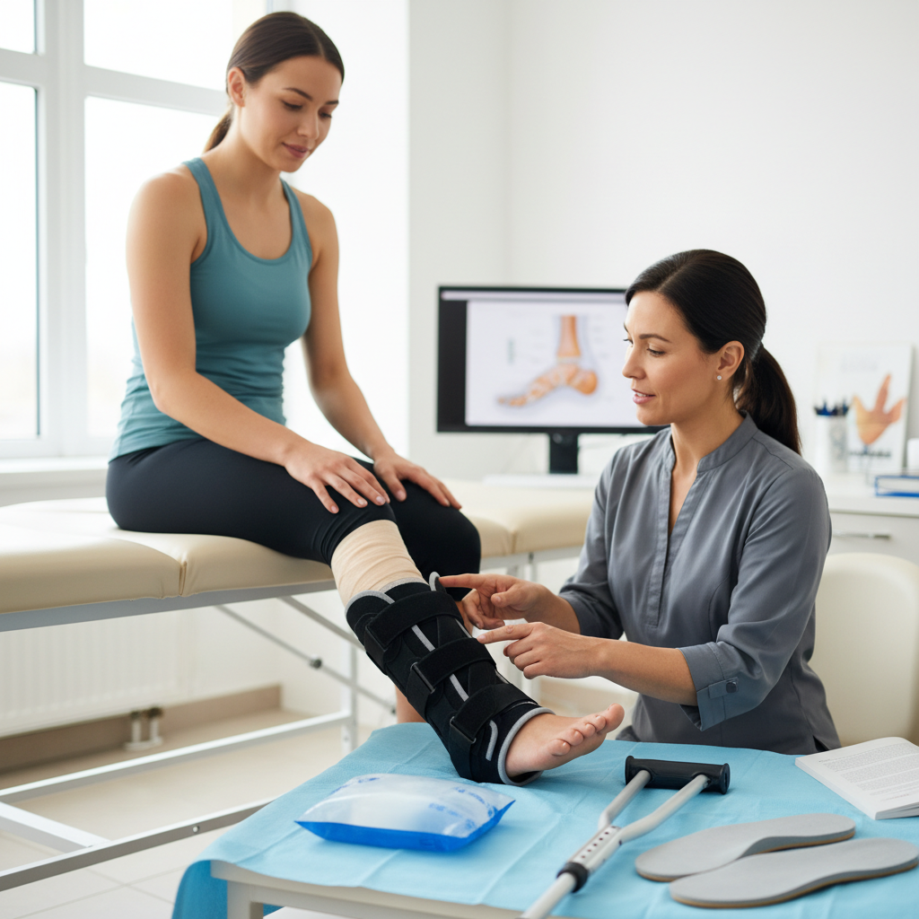
Stepping Toward Recovery: Your Guide to Healing an Ankle Sprain
This guide outlines the essential steps for recovering from an ankle sprain using the RICE method and bracing, while highlighting the clinical signs that indicate a need for professional podiatric care.
It starts with a single misstep on a curb or an awkward landing during a weekend game, followed by a sickening pop and a sudden surge of heat. An ankle sprain is more than just a temporary "tweak"—it is a complex soft-tissue injury that, if left unaddressed, can lead to a lifetime of instability and chronic pain.
What's actually happening
When you "roll" your ankle, you are experiencing an acute injury to the ligaments—the tough, fibrous bands of tissue that connect bones to other bones and provide stability to the joint. In the vast majority of cases, the foot turns inward (an inversion sprain), causing the ligaments on the outside of the ankle to stretch beyond their physiological limit.
The most commonly injured structure is the anterior talofibular ligament (ATFL). Depending on the force of the injury, these ligament fibers can undergo microscopic tearing (Grade I), a partial tear (Grade II), or a focal, complete rupture (Grade III). As soon as the fibers tear, your body initiates an inflammatory cascade. Blood flow increases to the area to deliver repair cells, which causes the hallmark swelling and bruising.
However, because ligaments have a relatively poor blood supply compared to muscles, they do not heal overnight. Without proper alignment and rehabilitation, the ligament may heal in a "lengthened" or lax position, which is why many people feel like their ankle is "giving out" months after the initial injury.
Signs and symptoms to watch for
While pain is the most obvious indicator, the specific way your ankle behaves in the hours following the injury can tell you a lot about the severity:
- Bruising and discoloration: This often appears as a "bruise necklace" around the base of the ankle and can even travel down into the toes due to gravity.
- Localized swelling: Swelling that appears immediately (within minutes) often indicates a more significant tear or even a fracture.
- Tenderness to touch: Feeling sharp pain when pressing directly on the bony bumps (malleoli) of the ankle.
- Instability: A sensation that the ankle is "wobbly" or unable to support your body weight.
- Limited range of motion: Feeling as though the joint is locked or too stiff to flex the foot upward or downward.
- The "Pop": Many patients report hearing or feeling a physical snap at the moment of impact.
Key takeaway: Not all ankle sprains are "just" sprains; the symptoms of a high-grade ligament tear often mimic those of a hairline fracture. Professional evaluation is essential to rule out bone damage.
Causes and risk factors
Ankle sprains occur when the joint is forced into an unnatural position, but several factors can make you more susceptible to this "tipping point."
Physical Activity and Environment Participating in sports that require pivoting, jumping, or lateral cutting—such as basketball, tennis, soccer, or trail running—places immense torque on the lateral ligaments. Uneven surfaces, ranging from cracked Irvine sidewalks to grassy fields, are classic culprits for sudden inversion.
Biomechanical Predisposition Some individuals have a "high arch" foot type (pes cavus). High arches naturally tilt the heel slightly inward, making it easier for the foot to roll over the outside edge. Similarly, if you have naturally "loose" joints (ligamentous laxity), your connective tissues may not provide the rigid "stopping point" needed to prevent a sprain.
Previous Injury History The single greatest risk factor for a sprain is a previous sprain that wasn't fully rehabilitated. When a ligament heals poorly, the proprioception—your brain's ability to sense where your foot is in space—is diminished. This lag in communication between the ankle and the brain means you won't be able to "catch" yourself the next time you stumble.
How it's diagnosed
A podiatrist begins by tracing the "mechanism of injury"—exactly how the foot turned and what you felt. Following a physical exam where the doctor checks for specific points of tenderness and joint laxity, imaging is often utilized to see beneath the surface.
- Digital X-rays: These are used primarily to rule out fractures. In Irvine, we often look for "avulsion fractures," where the ligament actually pulls a small piece of bone away from the fibula.
- Stress Views: Sometimes, the podiatrist gently moves the ankle under X-ray to see how much the joint "opens up," which indicates the degree of ligamentous laxity.
- Ultrasound: This allows for a real-time look at the soft tissues and can identify fluid buildup or a visible gap in the ligament fibers.
- MRI: Generally reserved for chronic cases or suspected Grade III tears, an MRI provides the most detailed view of the cartilage and ligaments.
Treatment options
The goal of treatment is twofold: reduce inflammation in the short term and restore mechanical stability in the long term.
Conservative care
The first 48 to 72 hours are critical. Most treatment plans begin with the R.I.C.E. protocol (Rest, Ice, Compression, and Elevation). However, modern sports medicine is shifting toward P.O.L.I.C.E. (Protection, Optimal Loading, Ice, Compression, Elevation), emphasizing that gentle, controlled movement is often better than total immobilization.
- Bracing and Splinting: A lace-up brace or a stirrup-style brace (like an Aircast) stabilizes the side-to-side motion of the ankle while still allowing you to move your foot up and down.
- Anti-inflammatory Medications: Non-steroidal anti-inflammatory drugs (NSAIDs) can help manage pain and reduce the swelling that inhibits healing.
- Physical Therapy: This is the cornerstone of recovery. Therapy focuses on strengthening the peroneal muscles (the muscles on the outside of the leg) to act as dynamic stabilizers for the weakened ligaments.
Advanced or minimally invasive options
If the ankle remains unstable or if there is internal scarring, we may look at modern regenerative or diagnostic techniques.
- Custom Orthotics: If your foot structure (like high arches) contributed to the sprain, custom-molded inserts can realign the heel and provide a wider, more stable "landing zone" for each step.
- Kinesiology Taping: This can help with lymphatic drainage (reducing swelling) and provide a sensory reminder to the brain to keep the ankle aligned.
Surgical options
Surgery is rarely the first choice for an initial sprain. However, for athletes with recurring "chronic ankle instability" or for complete ruptures that don't respond to therapy, surgical intervention may be necessary.
- The Broström Procedure: This is the "gold standard" for ankle stabilization. The surgeon shortens and tightens the overstretched ligaments to restore the joint's original tension.
- Ankle Arthroscopy: A "scope" is used to clean out loose fragments of bone or cartilage (debridement) that may be causing pain or catching sensations inside the joint.
Recovery and what to expect
Healing happens in phases, and skipping a phase is the most common reason for reinjury.
- Phase 1 (Days 1–7): The focus is on protection and reducing swelling. You may be in a walking boot or using a brace. The goal is to regain the ability to bear weight comfortably.
- Phase 2 (Weeks 2–4): You will begin gentle range-of-motion exercises. This is when "alphabet exercises" (tracing letters with your toes) and light stretching begin to break up scar tissue.
- Phase 3 (Weeks 4–8): This is the strengthening phase. You'll use resistance bands and perform "proprioceptive" drills, such as balancing on one leg.
- Phase 4 (Weeks 8–12+): Gradual return to sports. This involves "functional testing," where you practice cutting, jumping, and running in a controlled environment before returning to full competition.
Prevention and self-care tips
Preventing a second sprain is significantly easier than treating a chronic one.
- Invest in the right footwear: Replace athletic shoes every 300–500 miles. Shoes with a wide "outrigger" on the sole offer better lateral stability.
- Warm up dynamically: Instead of static stretching, use active movements like leg swings and ankle circles to wake up the nerves and muscles around the joint.
- Balance training: Spend two minutes a day standing on one foot while brushing your teeth. This "re-trains" the neurological connection between your ankle and your brain.
- Listen to the pain: If your ankle begins to ache toward the end of a workout, it is a sign that the stabilizing muscles are fatigued and the ligaments are taking the brunt of the force. Stop and rest.
- Taping or bracing for high-risk activities: If you are returning to a sport like volleyball or basketball, wearing a sleeve or brace for one full season post-injury provides an extra layer of mechanical insurance.
When to see a podiatrist
Self-treating with ice and rest is common, but several "red flags" indicate that the injury requires professional intervention from Dr. Eskander.
- You are unable to take four steps, even with a limp, immediately after the injury.
- The swelling does not begin to subside after 48 hours of elevation and icing.
- You feel "bone pain" directly on the medial (inner) or lateral (outer) bumps of the ankle.
- The ankle feels "loose" or gives way when you try to walk on a flat surface.
- You experience numbness or tingling in the foot or toes (indicating potential nerve involvement).
- The skin over the injury site is hot, red, or excessively tight and shiny.