
Sprained vs. Broken Ankle: How to Tell the Difference and When to Seek Help
Confused if your ankle injury is a simple sprain or a serious fracture? Learn the anatomical differences, key symptoms, and professional treatment options to ensure a safe recovery and prevent long-term instability.
That sudden pop or sickening crunch during a workout can leave you clutching your ankle in immediate uncertainty. While the pain of a severe sprain and a minor fracture often feel identical, the path to recovery for these two injuries is fundamentally different.
What's actually happening
To understand these injuries, you have to look at the structures holding your ankle together. A sprain occurs when the ligaments—the tough, fibrous bands of tissue that connect bone to bone—are stretched or torn. This usually happens when the foot rolls inward or outward beyond its normal range of motion.
An ankle fracture, on the other hand, involves a partial or complete break in one or more of the bones that form the ankle joint: the tibia (shinbone), the fibula (the smaller bone of the lower leg), or the talus. While a sprain involves soft tissue, a break involves the structural framework of your body. In some severe cases, a patient may experience both a fracture and a ligament tear simultaneously.
Signs and symptoms to watch for
Because both injuries involve inflammation and pain, it can be difficult to self-diagnose. However, certain symptoms are more characteristic of one over the other:
- Bruising and Swelling: Both cause this, but fractures often lead to rapid, localized swelling directly over the bone.
- Deformity: If the ankle looks crooked or out of place, a fracture or dislocation is likely.
- Sound: A 'pop' is common with sprains, while a 'crack' or 'grinding' sound often indicates a break.
- Weight Bearing: If you cannot take more than four steps without excruciating pain, the likelihood of a fracture increases significantly.
- Tenderness: Sprains usually hurt in the soft gaps between bones, whereas fractures are tender to the touch directly on the bony protrusions (malleoli).
Key Takeaway: Never assume an injury is 'just a sprain' simply because you can still walk on it; many minor fractures allow for limited weight-bearing but require professional stabilization to heal correctly.
Causes and risk factors
Most ankle injuries result from sudden rotational forces or direct impact. Common scenarios include tripping on an uneven sidewalk, landing awkwardly from a jump, or sports-related collisions.
Certain factors increase your vulnerability to these injuries:
- Previous Injuries: A history of untreated sprains can lead to chronic ankle instability.
- Improper Footwear: Wearing shoes that lack lateral support during high-impact activities.
- Bone Density: Conditions like osteoporosis can make the fibula more susceptible to stress fractures.
- Activity Level: Sudden increases in exercise intensity without proper conditioning.
How it's diagnosed
A professional evaluation begins with a physical exam to check for 'point tenderness' and range of motion. According to the Ottawa Ankle Rules, specific clinical markers help determine if imaging is necessary.
If a fracture is suspected, X-rays are the standard first step to visualize the bone. If the doctor suspects a high-grade ligament tear or a stress fracture that doesn't show up on a traditional X-ray, an MRI or CT scan may be ordered to provide a detailed view of the soft tissues and joint surfaces.
Treatment options
Conservative care
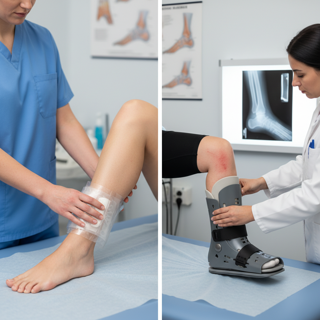
For mild to moderate sprains (Grade I and II), the R.I.C.E. method (Rest, Ice, Compression, Elevation) remains the gold standard. We may also utilize bracing or a walking boot to protect the ligaments while they knit back together. Physical therapy is essential here to restore proprioception—your brain's ability to sense the ankle's position.
Advanced/minimally invasive options
For stubborn sprains or minor non-displaced fractures, we may use functional bracing that allows for early protected movement. This prevents joint stiffness and muscle atrophy. In some cases, regenerative injections or specialized laser therapy may be discussed to accelerate the healing of soft tissue fibers.
Surgical options
Surgery is typically reserved for unstable fractures where the bones have shifted out of alignment (displaced) or for Grade III ligament tears that result in chronic instability. Surgeons may use small plates and screws to stabilize the bone or perform a ligament repair (Brostrom procedure) to tighten a chronically loose ankle.
Recovery and what to expect
Recovery timelines vary significantly based on the severity of the tissue damage. A mild sprain may feel better within 2 to 4 weeks, though full ligament strength can take months to return.
A broken ankle requires a much stricter timeline. Bone healing generally takes a minimum of 6 to 8 weeks of immobilization. Following this period, a structured rehabilitation program lasting 2 to 3 months is usually necessary to regain full strength, balance, and flexibility.
Prevention and self-care tips
- Strengthen your core and calves: Stronger muscles take the pressure off your joints.
- Warm up properly: Cold ligaments are less flexible and more prone to tearing.
- Replace worn-out shoes: Footwear that has lost its structural integrity increases the risk of rolling your ankle.
- Balance training: Practicing standing on one foot can improve the neuromuscular pathways that prevent falls.
When to see a podiatrist
- You heard a distinct cracking sound at the time of injury.
- The ankle joint appears deformed or misaligned.
- You experience numbness or tingling in the foot or toes.
- There is significant swelling that does not improve with 24 hours of icing and elevation.
- You are unable to put any weight on the foot without severe pain.
- The skin over the injury site is broken or severely bruised.