
Achilles Tendon Repair: A Comprehensive Guide to Your Recovery Timeline
Recovering from an Achilles tendon repair is a marathon, not a sprint. Learn about the stages of healing, from the first week of immobilization to returning to high-impact sports, and discover what to expect during each phase of your rehabilitation.
That sudden, sharp pop at the back of your ankle often feels like being kicked, but it marks the beginning of a significant healing journey. While Achilles tendon surgery is highly effective at restoring strength, the road to recovery requires patience, precision, and a structured approach to rehabilitation.
What's actually happening
The Achilles tendon is the largest and strongest tendon in the human body, connecting your calf muscles to your heel bone. When this tendon ruptures and requires surgical repair, a surgeon stitches the torn ends back together to restore the tension necessary for walking and jumping. Because tendons have a relatively limited blood supply compared to muscles, the body takes considerable time to weave new collagen fibers and regain structural integrity. This biological process cannot be rushed, as the newly repaired tissue must gradually transition from a fragile connection to a resilient powerhouse capable of supporting several times your body weight.
Signs and symptoms to watch for
Before and immediately after surgery, you may notice several key indicators of the injury and the healing process:
- A distinct 'popping' or 'snapping' sensation at the moment of injury.
- Significant swelling and bruising around the back of the ankle and heel.
- Inability to 'push off' the ground or stand on your tiptoes.
- A palpable gap or depression in the tendon area.
- Post-operative localized warmth and redness as the inflammatory healing phase begins.
Causes and risk factors
Most Achilles ruptures occur during 'stop-and-go' sports like basketball, tennis, or soccer. However, certain factors increase the likelihood of needing surgical intervention. A sudden increase in physical activity intensity, often referred to as 'weekend warrior' syndrome, is a primary driver. Additionally, certain medications like fluoroquinolone antibiotics or corticosteroid injections can weaken the tendon fibers. Anatomical factors, such as tight calf muscles or flat feet, also place excessive strain on the Achilles, making it more susceptible to failure under load.
How it's diagnosed
Diagnosis typically begins with a physical examination using the Thompson Test, where a provider squeezes the calf muscle to see if the foot flexes. If the foot remains still, a rupture is likely. To confirm the extent of the tear and plan the surgical approach, imaging is essential. An ultrasound can provide a real-time view of the tendon's movement, while an MRI (Magnetic Resonance Imaging) offers high-resolution details regarding the quality of the tissue and the exact location of the gap.
Treatment options
Conservative care
In some cases, particularly for sedentary patients or those with specific medical risks, non-surgical treatment involving functional bracing and early range-of-motion exercises may be considered. This relies on the body's ability to bridge the gap with scar tissue while the foot is held in a pointed position.
Advanced/minimally invasive options
Modern techniques now allow for percutaneous or minimally invasive repairs. These methods use smaller incisions and specialized instruments to pull the tendon ends together, which can lead to less scarring and a lower risk of wound complications compared to traditional open surgery.
Surgical options
Traditional open repair involves a larger incision to provide the surgeon with a direct view of the rupture. This is often preferred for complex tears or chronic ruptures where the tendon has retracted significantly. In some instances, a tendon transfer—using a nearby tendon to reinforce the repair—may be necessary to ensure long-term stability.
Key Takeaway: Success after Achilles surgery is 50% the skill of the surgeon and 50% the patient's commitment to a disciplined, phased physical therapy program.
Recovery and what to expect
The recovery timeline for an Achilles tendon repair is generally measured in months rather than weeks. While every patient is unique, the standard progression follows this trajectory:
- Weeks 0–2 (Protection): You will likely be in a cast or a specialized splint, strictly non-weight bearing. The focus is on wound healing and reducing swelling.
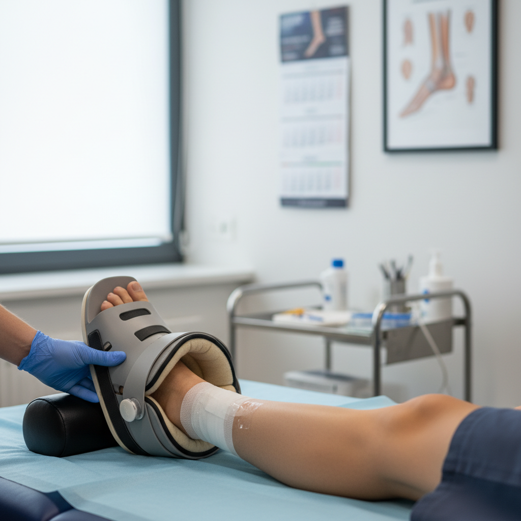
- Weeks 2–6 (Early Mobilization): You may transition to a walking boot with heel wedges. These wedges gradually decrease the angle of your foot, slowly stretching the tendon. Limited weight bearing often begins here.
- Weeks 6–12 (Strengthening): The boot is typically removed for daily activities. You will begin physical therapy to restore range of motion and start basic calf strengthening exercises.
- Months 3–6 (Functional Recovery): You will progress to more dynamic movements, such as light jogging and proprioception drills to improve balance.
- Months 6–12 (Return to Sport): Most patients return to high-impact athletics during this window, once strength is within 90% of the uninjured side.
Prevention and self-care tips
- Gradual progression: Never increase your workout intensity or duration by more than 10% per week.
- Eccentric strengthening: Incorporate 'heel drops' into your routine to build tendon resilience.
- Proper footwear: Replace athletic shoes every 300 to 500 miles to ensure adequate heel support.
- Hydration and nutrition: Ensure adequate intake of Vitamin C and collagen-supporting nutrients to aid tissue repair.
When to see a podiatrist
- If you experience a sudden 'pop' followed by an inability to walk.
- If you have persistent pain in the back of the heel that worsens with activity.
- If you notice new, sharp pain or a 'giving way' sensation during your post-op recovery.
- If the surgical site shows signs of infection, such as pus, increasing redness, or fever.
- If you experience calf pain or swelling that could indicate a blood clot.